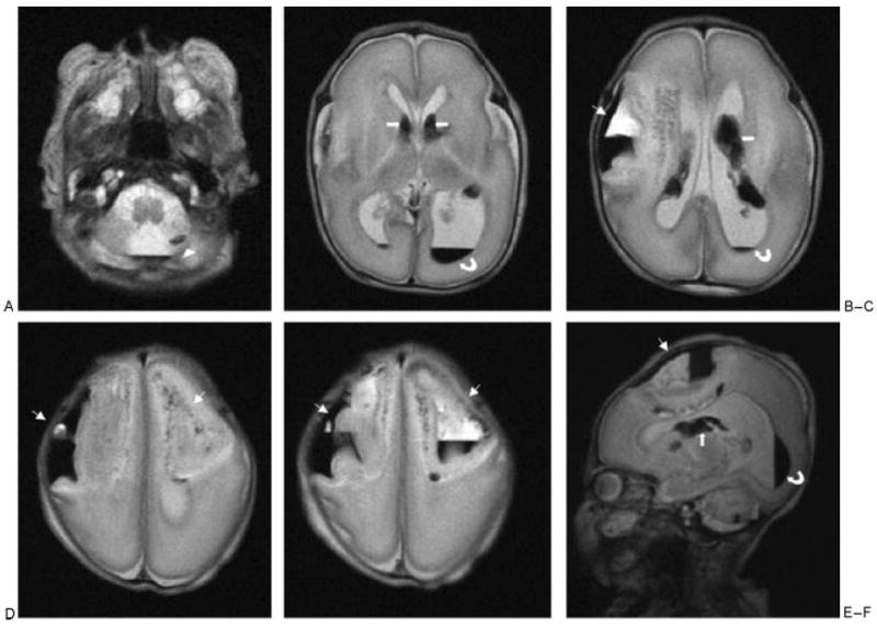
Figure 4.

Cerebral magnetic resonance imaging of patient 2, performed at day 6 of life ( weeks of corrected age). (A–E) Axial T2-weighted images showed these large bifrontal parenchymal hematomas (thin arrows), the left cerebellar hemorrhage (arrowheads), and bilateral intraventricular (curved arrows) and germinal matrix (thick arrows) hemorrhages. (F) Left sagittal T2-weighted image showed the left large frontal parenchymal hematomas (thin arrow), the left cerebellar hemorrhage, and left intraventricular (curved arrow) and germinal matrix (thick arrow) hemorrhages.
