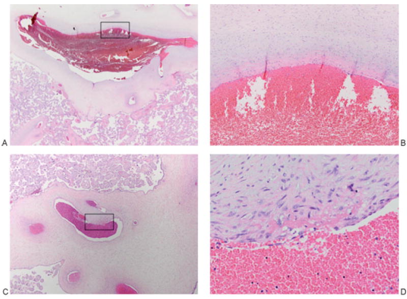
Figure 5.

Placenta of patient 2. (A, B) Organizing thrombus, chorionic plate vein: The vessel lumen is ectasic (left), and higher power shows it layered superiorly by a fibrin thrombus (B, represents boxed area in A). (A) 12.5×. (B) 200×. (C, D) Organizing thrombus, stem villous vessel: An ectasic lumen (left) at higher power shows a fibrin thrombus with early incorporation into the vessel wall (D, represents boxed area in C). (C) 40×. (D) 400×.
