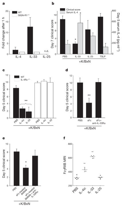
Figure 3. IL-33 triggers IL-4 anti-inflammatory activity.
a, Cytokine expression 1 h after IVIG administration in wild-type (black bars) or SIGN-R1−/− mice (white bars) determined by quantitative polymerase chain reaction (qPCR). n.d., not detected. b, K/BxN-treated wild-type mice received PBS, IL-33, IL-25, or TSLP. *P >0.05 determined by Tukey’s test. c, K/BxN-treated wild-type (black bars) or IL-4Rα−/− (white bars) mice received PBS, IL-4ic, or IL-33. **P >0.001 determined by Tukey’s test. d, hDC-SIGN+/SIGN-R1−/− mice received K/BxN sera, sFc and anti-IL-33Rα. **P >0.001 determined by Fisher LSD test. e, sFc-treated hDC-SIGN+ bone-marrow-derived macrophages were administered to wild-type mice, K/BxN- and anti-IL-33Rα-treated wild-type mice. Means and standard deviations are plotted; *P >0.05 determined by Tukey’s test. f, Individual mean fluorescence intensities (MFI) of bone marrow monocyte (CD11b+ Ly6G−) FcγRIIB surface expression 24 h after PBS, IL-4, IL-33, or IL-25 treatment by FACS. **P >0.01 determined by Tukey’s test.

