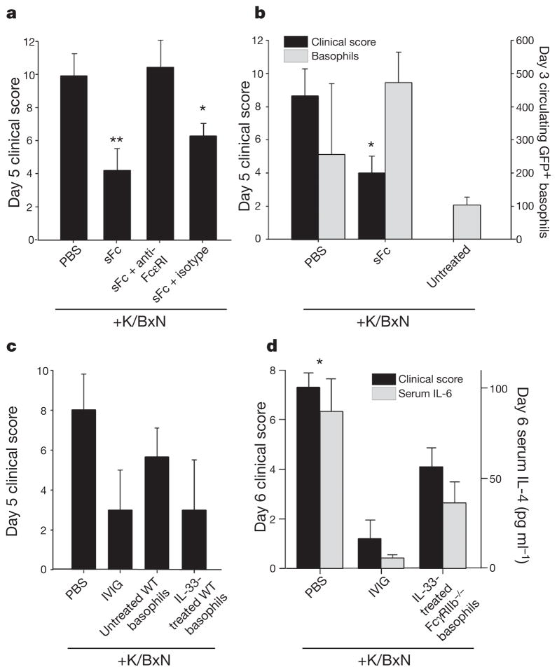
Figure 4. Anti-inflammatory activity mediated by basophils.
a, hDC-SIGN+/SIGN-R1−/− mice were administered K/BxN sera, sFc and anti-FcεRI or an isotype control. **P >0.001, *P >0.05 determined by Fisher LSD test. b, 4get mice were administered K/BxN sera and sFc. Circulating IL-4+ basophils (grey bars, DX5+ FcεRI+ GFP+) and clinical scores (black bars) are plotted. *P >0.05 determined by Tukey’s test. c, PBS or IL-33-treated basophils (DX5+ FcεRI+ c-Kit−) were administered to K/BxN-treated wild-type mice. Control mice received PBS or IVIG. d, Basophils from IL-33-treated FcγRIIB−/− mice were administered to K/BxN-treated wild-type recipients. Clinical scores (black) and serum IL-6 levels (grey) are plotted. Means and standard deviations are plotted;*P >0.05 determined by Mann–Whitney’s U test.

