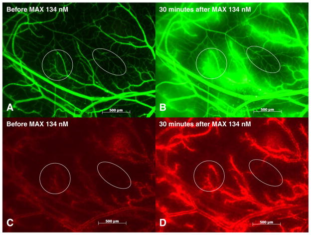
Fig. 1.
Maxadilan induced plasma leakage and leukocyte accumulation. Fluorescent light images of the observed area of 5 mm3 in one hamster cheek pouch preparation showing plasma leakage (a and b) and leukocyte accumulation (c and d) at 30 min after topical application of maxadilan 134 nM. Plasma leakage from postcapillary and larger venules is shown in (b). Leukocyte accumulation in postcapillary and larger venules is shown in (d). Circles include an area with postcapillary venules and ellipses include a different area with terminal arterioles and capillaries.

