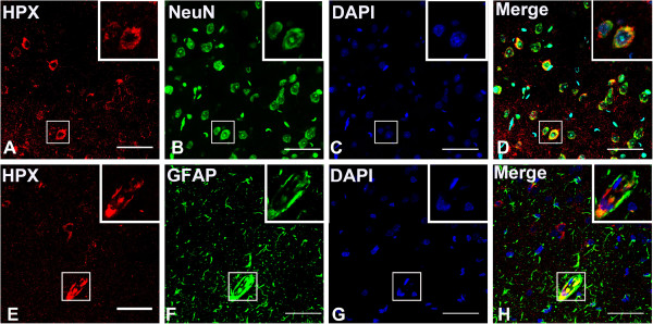
Figure 3.

Localication of HPX in neurons and astrocytes in the cerebral cortex. HPX was stained in red and NeuN/GFAP was stained in green. Note that HPX staining was strong in the cytoplasm of neurons but was weak in the nucleus (A-D,E-H), and the immunoreactive astrocytes were mainly located near the blood vessels (E-H). Scale bars = 40 μm.
