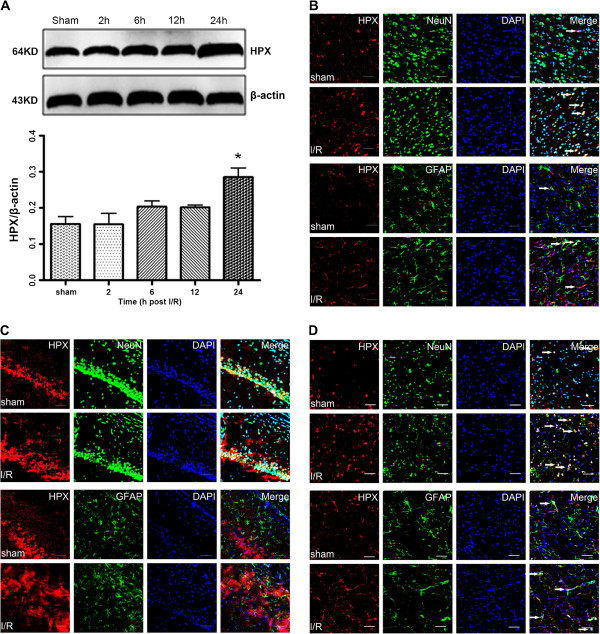
Figure 4.
HPX expression was evaluated in the ischemic penumbra at different time points following reperfusion. A, Western blotting of the HPX expression in sham and MCAO rats at 2 h, 12 h and 24 h after reperfusion. The expression of β-actin served as a loading control of the protein samples. The relative density of HPX protein to β-actin protein expression among groups was plotted. Data represent mean ± SEM. *P< 0.05 vs. Sham group. (n =4 for each group) Double immunofluorescence staining of HPX and NeuN/GFAP indicated that HPX expression in the cortex B, hippocampus C, and striatum D, were notably increased in the I/R group. Scale bars = 20 μm.

