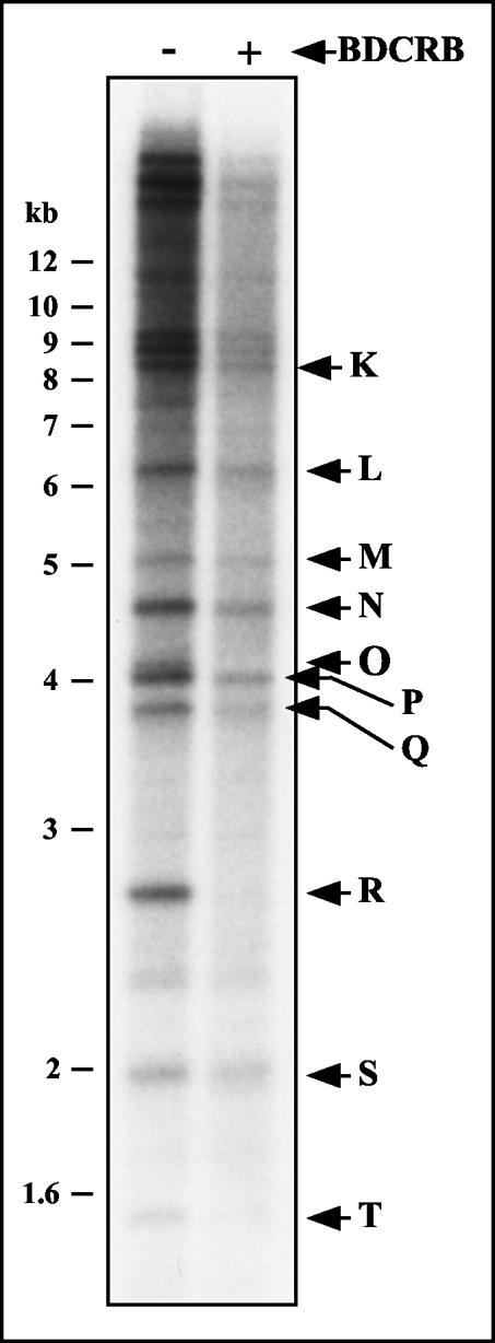
FIG. 7.
Restriction fragment pattern analysis of GPCMV genomes formed in the presence of BDCRB. Duplicate cultures were infected with GPCMV at an MOI of 3. The culture media was replaced at 72 h p.i. with media alone or media containing 50 μM BDCRB. Viral DNA was labeled between 80 and 96 h p.i. by the addition of [32P]orthophosphate to the media of both cultures. Intracellular DNA was prepared at 96 h p.i. and separated by FIGE. Intracellular 230-kb GPCMV DNA was excised in agarose blocks, extracted from the agarose, and digested with HindIII. The resulting restriction fragments were separated by agarose electrophoresis, transferred to a nylon membrane, and autoradiographed. Arrows indicate the locations of GPCMV HindIII restriction fragments surmised from their published molecular weights (32).

