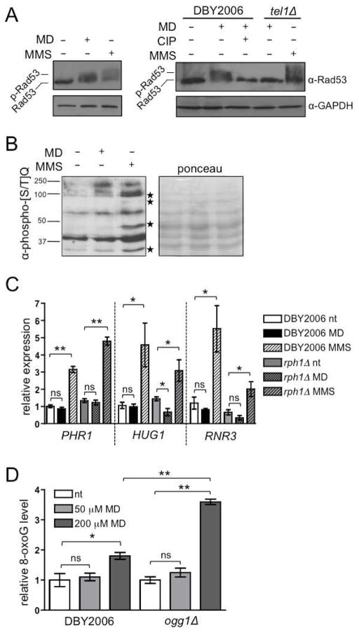
Figure 4. mtROS Activate a Non-canonical, Tel1p-dependent DNA Damage Response.
(A) Left: western blot analysis of Rad53p phosphorylation in the wild-type treated with 50 μM menadione (MD+), 0.01% methylmethane sulfate (MMS+) or ethanol (−) for 30 minutes. Right: wild-type and tel1Δ strains treated with menadione, MMS, ethanol and/or calf intestinal phosphatase (CIP).
(B) Protein phosphorylation at [S/T]Q sites in response to 30 minute treatment with 50 μM menadione or 0.01% MMS. Molecular weights (kDa) are indicated on the left. Starred bands indicate proteins that are phosphorylated in response to MMS treatment but not menadione treatment.
(C) RT-PCR of transcripts induced by DNA damage. Wild-type and rph1Δ were treated with 50 μM menadione (MD), 0.01% MMS, or vehicle (nt) for 30 minutes in exponential phase. See also Table S2.
(D) Quantification of 8-hydroxyguanosine (8-oxoG) formation in wild-type and ogg1Δ. Cells were treated for 30 minutes prior to DNA extraction. Values were normalized to untreated wild-type, which was set to one. Data points represent the mean of three biological replicates inoculated from single colonies ±SEM. “ns” designates p>0.05, * designates p≤0.05, ** designates p≤0.01. See also Figure S3.

