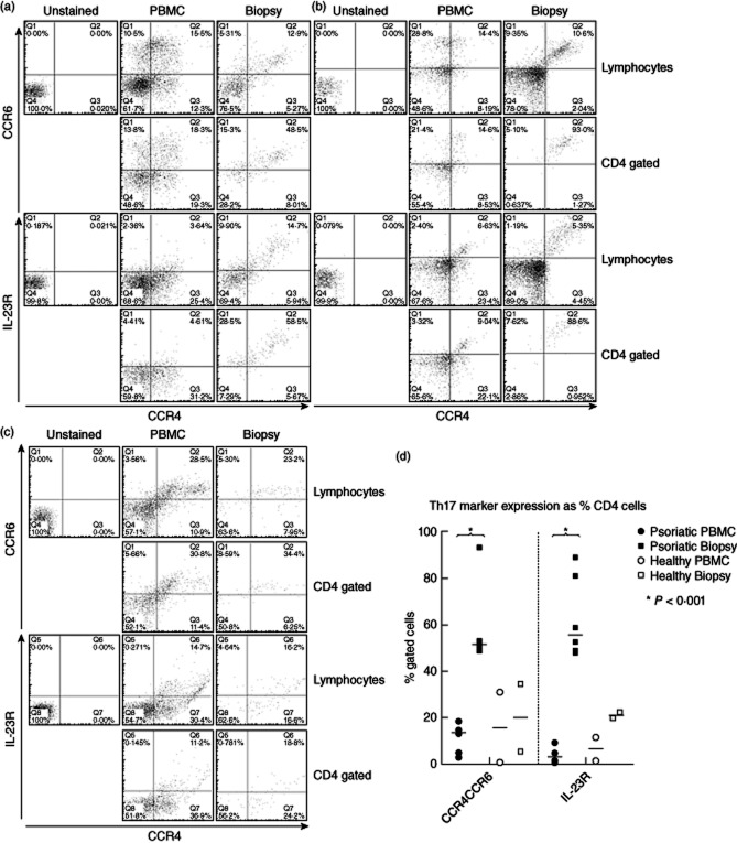
Figure 1.
CD4+ lymphocytes expressing T helper type 17 (Th17) markers CCR4 and CCR6 or interleukin (IL)-23R are greatly enriched in psoriatic lesions compared with peripheral blood. (a,b) Representative flow cytometric analyses from two patients and (c) from a healthy control. For each set, the expression of CCR4 and CCR6 (upper set) or CCR4 and IL-23R (lower set) are illustrated for paired samples of peripheral blood mononuclear cells (PBMC) (middle) and lesional cells (left), gated on the lymphocyte population (upper pairs), or CD4+CD3+ cells (lower pairs). Th17 cells stain CCR4+CCR6+ or CCR4+IL-23R+, (upper right quadrants). (d) Summarizes data from analyses of CD4+ cells from lesional biopsies and blood of psoriatic patients (n = 6) and healthy controls (n = 2), demonstrating the percentages of CD4+ cells that express Th17 markers (bar indicates median value; *P < 0·001, Kruskal–Wallis test).

