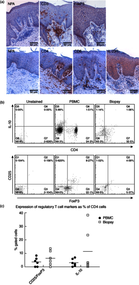
Figure 4.

Natural and induced regulatory T cells (Tregs) are present in psoriatic plaques, but while the frequency of the natural type is similar to that in peripheral blood, induced Tregs can be enriched in lesions from some patients. (a) Immunohistochemistry slides showing that CD4+ lymphocytes, forkhead box protein 3 (FoxP3+) cells and interleukin (IL)-23R+ cells are all found in both the dermis and epidermis of psoriatic lesions. (b) A set of representative flow cytometric data from psoriatic patients are shown, demonstrating the numbers of IL-10+-induced T regulatory 1(Tr1) phenotype (upper) and CD25hiFoxP3+ cells within the CD4+ lymphocyte (lower) populations from peripheral blood mononuclear cells (PBMC) (middle) and paired lesional biopsies (right). (c) Summarizes data from analyses of CD4+ cells from lesional biopsies and blood of psoriatic patients (n = 6), demonstrating the percentages of CD4+ cells that express Treg markers (bar indicates median value).
