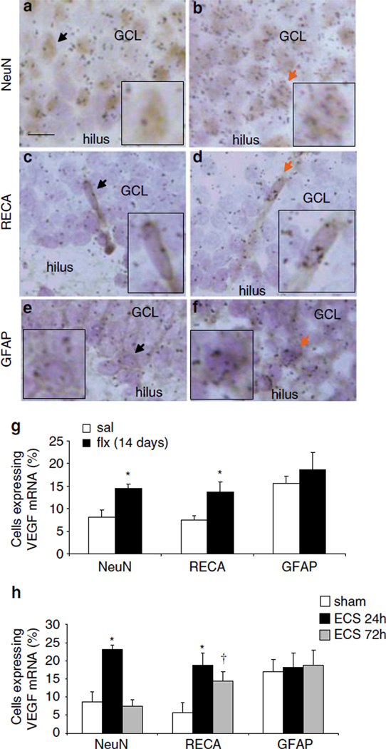
Figure 6.
VEGF mRNA is expressed by neurons and endothelial cells: regulation by ECS and fluoxetine treatment. Immunohistochemistry with phenotypic markers of neurons (NeuN) or endoethelial cells (RECA) followed by in situ hybridization for VEGF mRNA (black grains). (a, b) Representative images of the GCL cells immunolabeled with either NeuN (a, b), RECA (c, d) (brown) or GFAP antibody (e, f) counterstained with cresyl violet (blue); Scalebar = 10 µm. Sections are from control (a, c, e) or ECS (b, d, f). In all images, arrows indicate NeuN-, RECA-, or GFAP-positive cells expressing VEGF mRNA. Background was defined as 3 grains per cell, which was the approximate number of grains counted over a comparable area in a cell-sparse region such as the hilus. Inset images highlight the arrow-marked cell at 200% magnification. (g) Quantification of the influence of fluoxetine (FLX) on the number of cells expressing VEGF mRNA above background levels the in the GCL (NeuN: F(1,6) = 10.000, P<0.05; RECA: F(1,6) = 6.818, P<0.05). (h) Quantification of the influence of ECS on the number of cells expressing VEGF mRNA in the GCL (NeuN: F(2,9) = 19.966; P<0.01; RECA: F(2,9) = 5.304; P<0.05). *P<0.05, †P = 0.06 compared with saline or sham ECS.

