
Fig. 1.
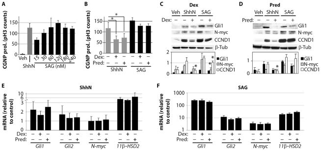
SAG antagonizes GC effects on cultured primary CGNPs. (A) CGNP proliferation (prol.) stimulated by various SAG concentrations (15 to 240 nM) compared with ShhN (3 μg/ml) and vehicle (Veh) after 24 hours in vitro (n = 4). (B) Effects of dexamethasone (Dex) (40 μM) and prednisolone (Pred) (120 nM) on ShhN-induced (P < 0.001, ANOVA with Tukey’s post hoc; n = 5) and 120 nM SAG–induced CGNP cultures (no significant change). (C and D) Western blots of protein lysates prepared from CGNPs treated with vehicle, ShhN, or 120 nM SAG in the presence or absence of 40 μM dexamethasone (C) [Gli1: P < 0.005, ANOVA; N-myc: P < 0.02, ANOVA (Tukey’s post hoc: SAG versus SAG + dexamethasone, P = 0.02); CCND1: P < 0.005, ANOVA (Tukey’s post hoc: ShhN versus ShhN + dexamethasone, P < 0.0001; SAG versus SAG + dexamethasone, P = 0.05)] or 120 nM prednisolone (D) [Gli1: P < 0.03, ANOVA; N-myc: P < 0.001, ANOVA; CCND1: P < 0.001, ANOVA (Tukey’s post hoc: ShhN versus ShhN + prednisolone, P = 0.04)] for 24 hours (n = 3). Signal intensity of the bands is illustrated in the histograms below. (E and F) Total RNA was isolated from CGNP cultures treated with vehicle, ShhN (E), or 120 nM SAG (F) after 24 hours in the presence or absence of 40 μM dexamethasone or 120 nM prednisolone (n = 3; no significant changes). Asterisks indicate significant changes using Tukey’s post hoc test.