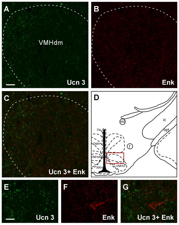
Fig. 11.
Fluorescent confocal images showing Ucn 3 (A) and Enk (B) immunostaining in the VMH. The location of the images was indicated by the red box in D. C: Merged image of A and B showing no significant colocalization of Ucn 3 and Enk in the VMH. E–G: High magnification of the dorsomedial part of the VMH showing the staining of Ucn 3 (E) and Enk (F). Merged image (G) of E and F further illustrates little colocalization of the two neurosubstrates in this brain area. 3V: Third ventricle, ARH: Arcuate nucleus of hypothalamus, DMH: Dorsomedial nucleus of hypothalamus, f: Fornix, ic: Internal capsule, mt: Mammillothalamic tract, opt: Optic tract, VMHdm and VMHvl: Dorsomedial part (VMHdm) and ventrolateral part (VMHvl) of the ventromedial nucleus of hypothalamus. Scale bar: 50 μm (A), 20 μm (E).

