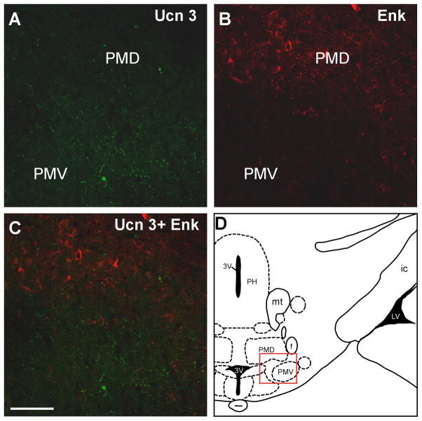
Fig. 12.
Fluoresecnt confocal images showing Ucn 3 (A) and Enk (B) in the premammillary area indicated by the red box in D. C: Merged image of A and B indicates that Ucn 3 immunoreacitivty was found mostly in the PMV where as prominent Enk staining was observed in the PMD and thus no significant colocalization of Ucn 3 and Enk was found in these brain regions. 3V: Third ventricle, f: Fornix, ic: Internal capsule, LV: Lateral ventricle, mt: Mammillothalamic tract, PH: Posterior hypothalamus, PMD: Dorsal premammillary nucleus, PMV: Ventral premammillary nucleus. Scale bar: 50 μm.

