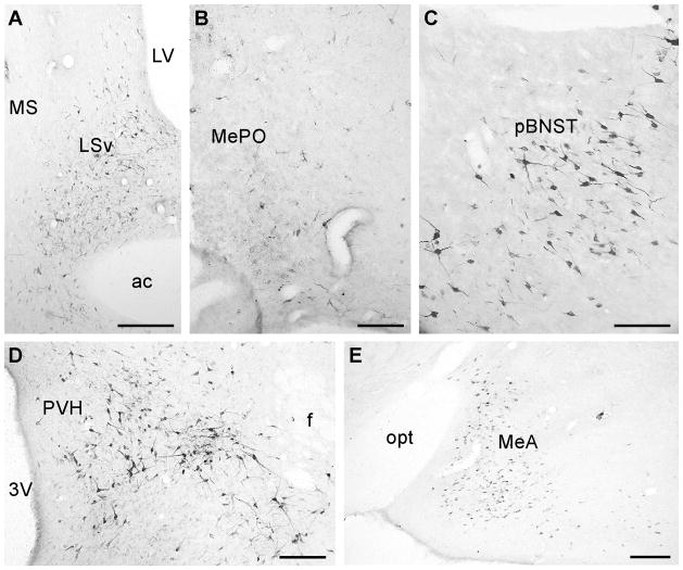
Fig. 2.
Representative photomicrographs showing FG-positive cells in the LSv (a), MePO (b), pBNST (c), perifornical hypothalamic area (d) and MeA (e). 3V: Third ventricle, ac: Anterior commissure, f: Fornix, LSv: Lateral septum, ventral part, LV: Lateral ventricle, MeA: Medial amygdala, MePO: Median preoptic nucleus, MS: Medial septum, pBNST: posterior part of the bed nucleus of stria terminalis, PVH: Paraventricular nucleus of hypothalamus, opt: Optical tract. Scale bar = 250 μm (a, e), 100 μm (b, c, d).

