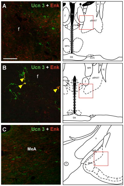
Fig. 9.
a: Double-label immunofluorescent staining of Ucn 3 (green) and Enk (red) in the posterior part of the BNST (A), the PVHap (B) and in the medial amygdala (C). The location of each image is indicated by red box in the respective drawing. Note that both Ucn 3 and Enk-positive cells and fibers are evident in these areas, but double-label cells or fibers were rarely observed. A few double-labeled cells and fibers (indicated by yellow arrowheads) were observed in the PVHap (B). 3V: Third ventricle, f: Fornix, ic: Internal capsule, MPN: Medial preoptic nucleus, LV: Lateral ventricle, ox: Optic chiasm, MeA: Medial amygdala, pBNST: posterior part of the bed nucleus of stria terminalis, PVHap: Anterior parvicellular part of the paraventricular nucleus of hypothalamus, SCN: Suprachiasmatic nucleus of hypothalamus, SON: Supraoptic nucleus of hypothalamus, sm: Stria medullaris of thalamus, st: Stria terminalis. Scale bar: 50 μm.

