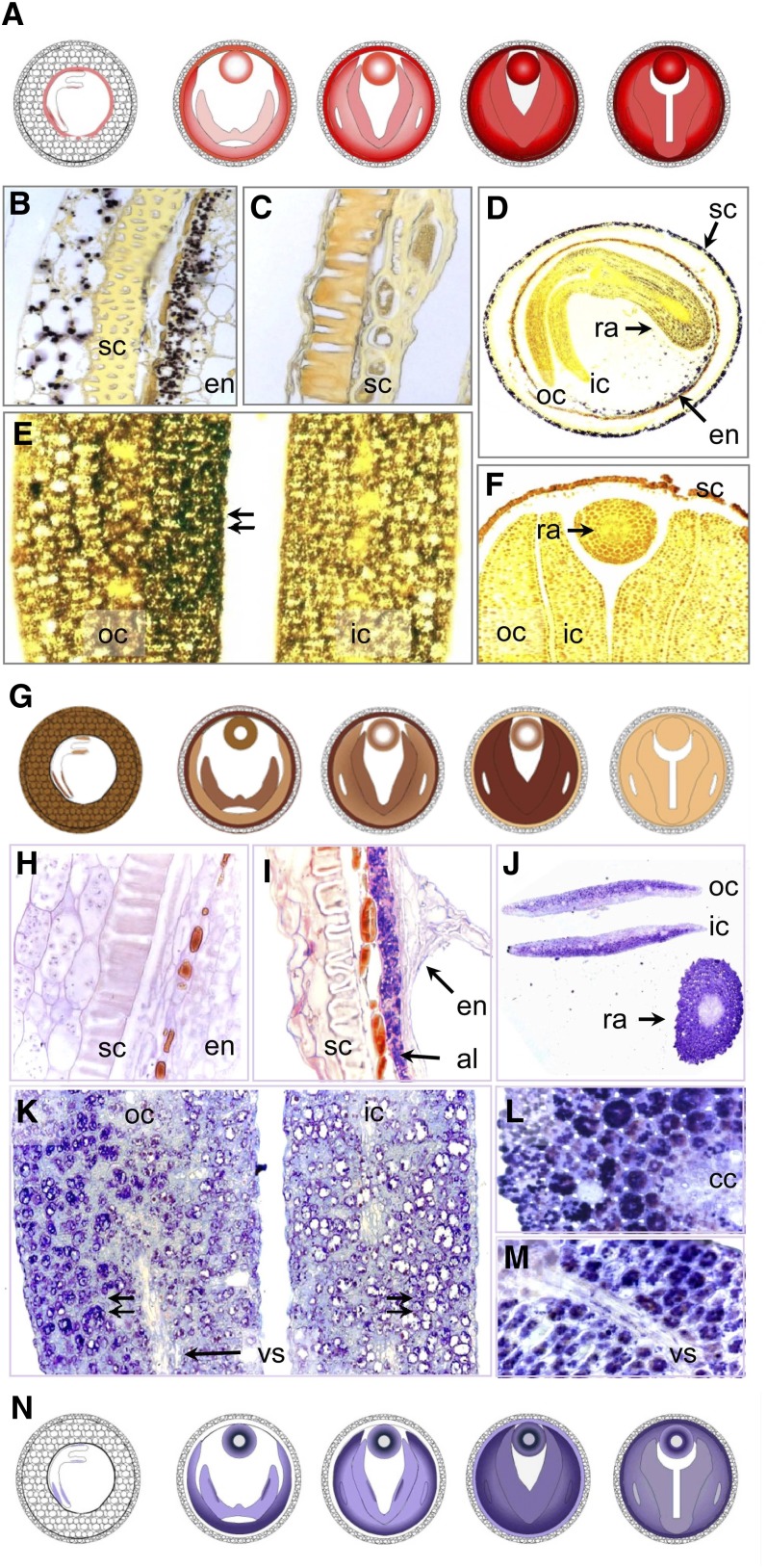
Figure 5.
Colocalization of Oleosin, Cruciferin, and Starch in the Developing Oilseed Rape Seed.
(A) Schematic representation of oleosin distribution (for details, see Supplemental Figure 2 online).
(B) to (F) Starch deposition (iodine staining).
(B) Starch grains are visible within the seed coat and endosperm during early storage stage.
(C) Starch is no longer detectable in the seed coat during the later stages.
(D) A cross section through the seed shows the presence of starch in the seed coat, endosperm, and embryo, visible as a gradient from the radicle toward the cotyledon. The outer cotyledon is more heavily stained than the inner one.
(E) A gradient in starch content within the outer cotyledon (cf. with the protein gradient shown in [K]).
(F) A cross section through the mature seed shows the complete absence of starch.
(G) Schematic representation of starch distribution.
(H) to (M) Cruciferin deposition (immunostaining).
(H) A cross-section through the seed coat and endosperm during the early storage stage shows no cruciferin to be present;
(I) The mid storage stage endosperm contains plenty of cruciferin.
(J) A cross section of the young seed demonstrates the presence of cruciferin in the embryo and cotyledons. A gradient in concentration is visible in both radicle and cotyledons.
(K) A cross section through the cotyledons during the mid storage stage shows a cruciferin gradient within the outer cotyledon. The storage vacuoles in the abaxial region are full of protein (double arrow left), those closer to the seed's interior are less filled, while those in inner cotyledon are empty (double arrow right).
(L) Cortical tissue of radicle showing cells completely full of cruciferin.
(M) The cotyledonary vascular tissue is relatively free of cruciferin.
(N) Schematic representation of cruciferin distribution.
al, aleurone layer; cc, central cylinder; en, endosperm; ic, inner cotyledon; oc, outer cotyledon; ra, radicle; sc, seed coat; vs, vascular tissue.

