Figure 1.
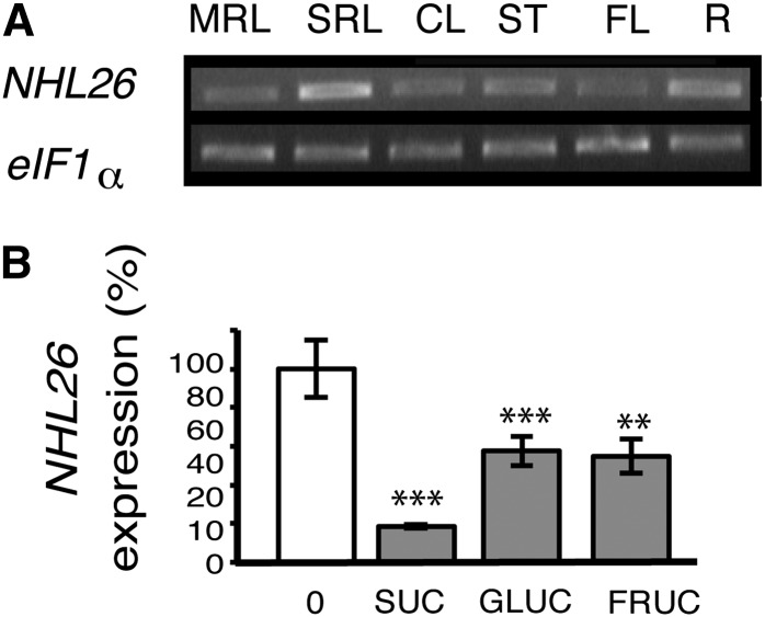
Pattern of Expression of NHL26.
(A) Expression of NHL26 in various organs. The accumulation of NHL26 RNA was analyzed by RT-PCR. EiF1α was used as an internal quantitative control. RT-PCR products were detected by ethidium bromide staining of the agarose gels used for electrophoresis of the PCR products. CL, cauline leaves; FL, flowers; MRL, mature rosette leaves; R, roots; SRL, senescent rosette leaves; ST, stem leaves.
(B) Transcriptional regulation of NHL26 by sugars. The expression of NHL26 was analyzed by qRT-PCR on 7-d-old Arabidopsis seedlings treated for 6 h with 10 mM Suc, Glc, or Fru. Expression levels are indicated as percentages of the values obtained for the untreated samples (0 mM). The levels of NHL26 transcript accumulation were normalized relative to levels of APT RNA. Asterisks indicate values that are significantly different, as assessed in a t test (**P < 0.02; ***P < 0.01). Error bars indicate the se (n = 6).

