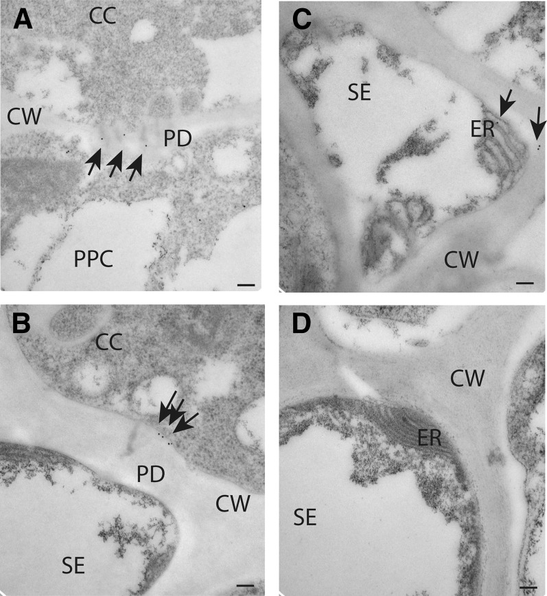
Figure 10.
Localization of NHL26 in PD and in the SE Reticulum.
NHL26 was localized by immunogold labeling and transmission electron microscopy. Arrows indicate gold particles conjugated to an anti-GFP polyclonal antibody. Bars = 100 nm.
(A) to (C) Detection of the NHL26-GFP fusion protein with an anti-GFP antibody in phloem cells in pNHL:NHL-GFP Arabidopsis plants.
(A) Details of localization between phloem parenchyma and CCs, showing localization to PD.
(B) Details of localization between CCs and SEs, showing localization to PD.
(C) Details of localization in SEs, showing localization to the SE reticulum.
(D) Negative control for immunogold labeling.

