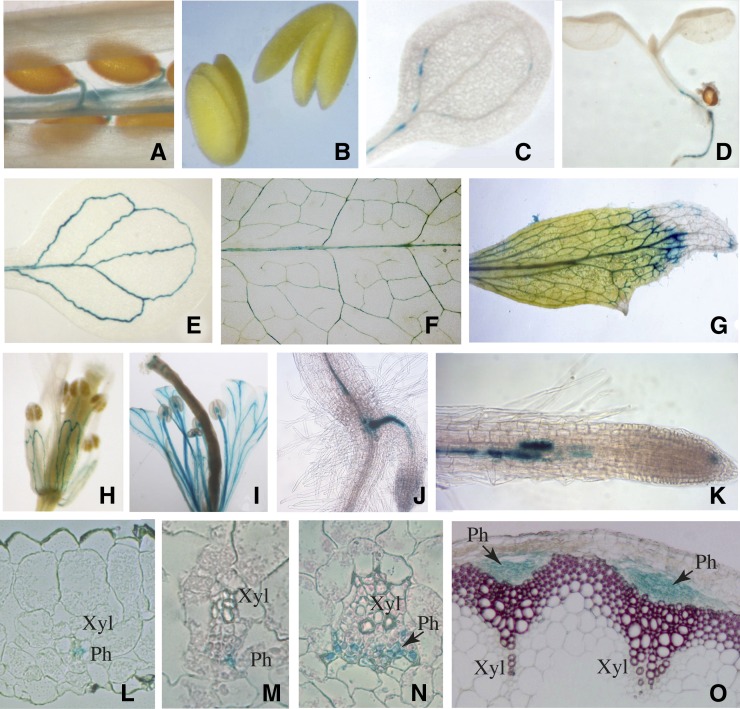
Figure 2.
Organ and Tissue Localization of NHL26 Expression.
Representative GUS staining of pNHL:GUS-expressing plants. (A) Silique; (B) mature embryos; (C) cotyledon 3 d after germination; (D) 8-d-old seedling; (E) cotyledon 8 d after germination; (F) mature rosette leaf; (G) senescent cauline leaf; (H) and (I) flowers observed before and after pollination, respectively; (J) hypocotyl-root junction with emerging adventitious root; (K) primary root; (L) transverse thin section of minor vein from a mature leaf; (M) and (N) transverse thin section of a secondary vein from a mature leaf; (O) transverse thin section of floral stem (the lignin was stained with phloroglucinol). Arrows, phloem; Ph, phloem; Xyl, xylem.

