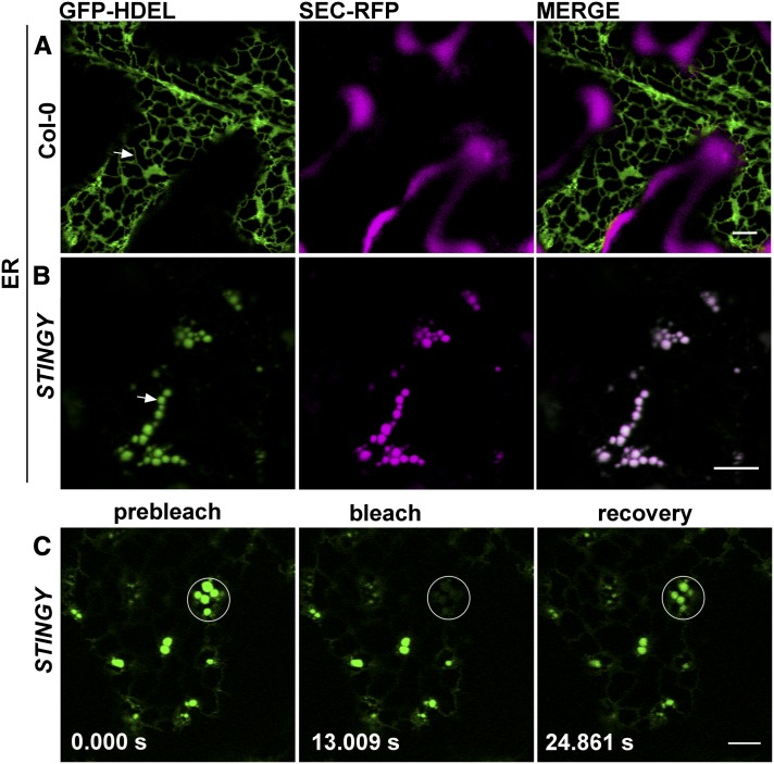
Figure 2.
SEC-RFP Distribution in STINGY Coincides with That of a Soluble ER Marker into Globular Structures That Are Interconnected.
(A) Confocal optical section of a SEC-RFP cotyledon epidermal cell expressing the ER marker GFP-HDEL, which localizes to the lumen of the ER and defines the ER network.
(B) Confocal optical section of STINGY cotyledon epidermal cells expressing the GFP-HDEL marker showing an overlap of the RFP and GFP signals in the globular structures of the mutant.
(C) Photobleaching of globular structures and recovery of fluorescence indicate that the globular structures are not isolated. Time of frame acquisition is indicated on individual panels in seconds.
Bars = 5 μm.

