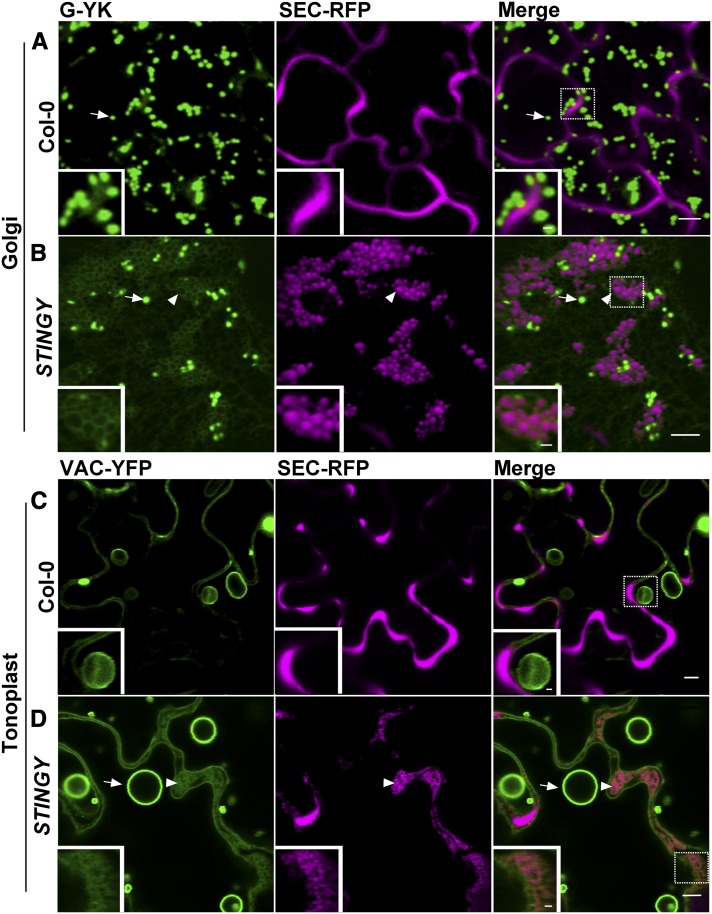
Figure 3.
The STINGY Globular Structures Are Contained in a Modified ER Membrane in Which Golgi and Tonoplast Proteins Are Partially Retained.
(A) and (B) Confocal optical section of Col0/SEC-RFP control coexpressing the Golgi marker G-YK showing that the marker is localized to the Golgi apparatus ([A], arrow), as expected. However, STINGY/SEC-RFP coexpressing G-YK shows partial accumulation of the Golgi marker into a modified ER network enwrapping the globular STINGY structures ([B], arrowhead and inset).
(C) and (D) Confocal images of Col-0/SEC-RFP coexpressing the tonoplast marker VAC-YFP showing tonoplast (arrow) and bulb structures (arrowhead). In the STINGY mutant, the marker partially accumulated in circular structures (arrowhead and inset).
Bars = 5 μm; bars in the insets = 1 μm. Insets show enlargements of the boxed regions.

