Figure 1.
VLN2 and VLN5 Decorate Filamentous Structures in Pollen Tubes.
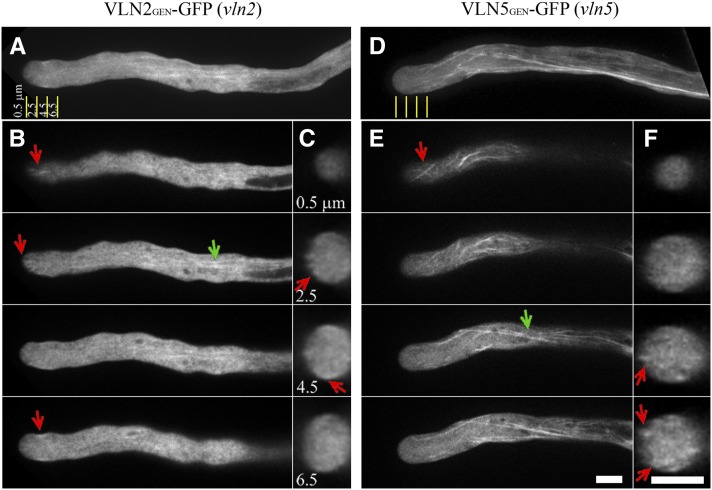
VLN2GEN-GFP or VLN5GEN-GFP was transformed into vln2 or vln5 lines, respectively, to assess the localization of VLN2 or VLN5 in pollen tubes.
(A) to (C) VLN2 localization in the pollen tube.
(D) to (F) VLN5 localization in the pollen tube.
(A) The projection showed that VLN2 decorates filamentous structures in the shank as well as the subapex and apex. Yellow lines indicate distances distal from the apex, and the transverse sections were captured at the positions indicated in (C).
(B) Selected single optical sections. Red arrows indicate VLN2-decorated filamentous structures at the apex and subapex. Green arrow indicates VLN2-decorated filamentous structures in the shank.
(C) Transverse sections of the pollen tube taken at the positions indicated by the yellow lines at specific distances distal from the apex. Red arrows indicate VLN2-GFP fluorescence in the cortical region.
(D) Projections revealed that VLN5 decorates filamentous structures throughout the pollen tube. Yellow lines indicate distances distal from the apex identical to those shown in (A).
(E) Selected single optical sections. Red and green arrows indicate VLN5-decorated filamentous structures at the tip and shank, respectively. Bar = 5 μm in (E) for (A), (B), (D), and (E).
(F) Transverse sections of the pollen tube taken at the positions indicated by the yellow lines at specific distances distal from the apex. Red arrows indicate VLN5-GFP fluorescence in the cortical region. Bar = 5 μm in (F) for (C) and (F).

