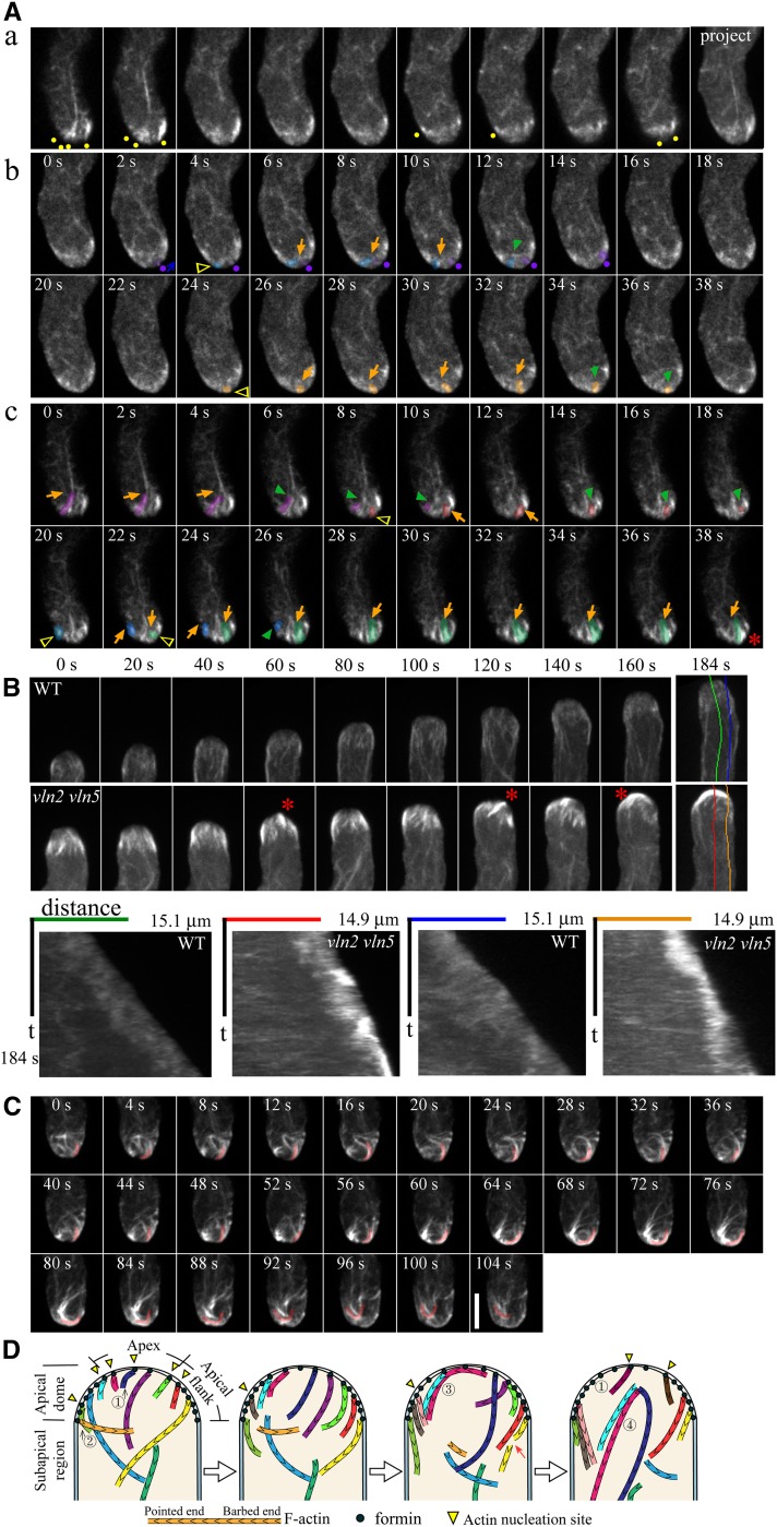
Figure 2.
Downregulation of VLN2 and VLN5 Causes Filamentous Actin to Accumulate at Pollen Tube Tips.
(A) Visualization of actin filaments in the apical region of vln2 vln5 pollen tubes. (a) z-series optical sections and projections revealed that actin filaments are present at the apex and apical flank of the vln2 vln5 pollen tube. Yellow dots indicate the positions of actin filaments close to the membrane. (b) Time-lapse images of actin filaments in the medial section of the apical dome of a vln2 vln5 pollen tube. Three actin filaments with different behaviors were pseudocolored purple, blue, and yellow. Yellow triangles indicate the origination sites of actin filaments, and purple dots indicate the position of anchorage of an actin filament that moves from the apex to the apical flank. Blue arrow indicates the moving direction of actin filaments. Orange and green arrows indicate growing and shrinking actin filaments, respectively. See Supplemental Movie 5 online for the entire series. (c) Time-lapse images of actin filaments in the cortical region of the apical dome of a vln2 vln5 pollen tube. Four actin filaments with distinct behaviors were pseudocolored purple, orange, blue, and green. Yellow triangles indicate the association sites of actin filaments with the membrane. Orange and green arrows indicate growing and shrinking actin filaments, respectively. The red asterisk indicates an accumulation of actin filaments. See Supplemental Movie 6 online for the entire series.
(B) Actin filaments accumulate at the apex and the apical flank of vln2 vln5 pollen tubes. The top panel shows time-lapse images of actin filaments in a wild-type (WT) pollen tube (see Supplemental Movie 7 online for the entire series) and a vln2 vln5 pollen tube (see Supplemental Movie 8 online for the entire series). Red asterisks indicate the position of actin filament accumulation. Kymographs were plotted based on lines parallel to the growth axes of pollen tubes, demonstrating that actin filaments accumulate in vln2 vln5 pollen tube tips.
(C) Time-lapse images of actin filaments from another vln2 vln5 pollen tube showed that actin filaments generated at the apical membrane were not severed and turned over. Consequently, they accumulated and intertwined near the membrane. They then departed from the membrane as a unit (see the represented pink pseudocolored filament), reducing the amount of actin filaments at pollen tube tips. Bar = 5 μm for images in (A) to (C).
(D) Schematic diagram of actin filament dynamics within the apical and subapical regions of vln2 vln5 pollen tubes. Similar to wild-type pollen tubes, nucleation of actin filaments is probably mediated by membrane-anchored formin(s). However, unlike wild-type pollen tubes, actin filaments accumulated and intertwined near the membrane within the apical dome of vln2 vln5 pollen tubes (see 3 marked actin filaments). Actin filaments are able to be nucleated on membrane at both the apex (see 1 marked actin filaments) and the apical flanks (see 2 marked actin filaments). Some intertwined actin structures leave the membrane as a unit via an unknown mechanism (see 4 marked actin filaments). Red arrow indicates the severing event.

