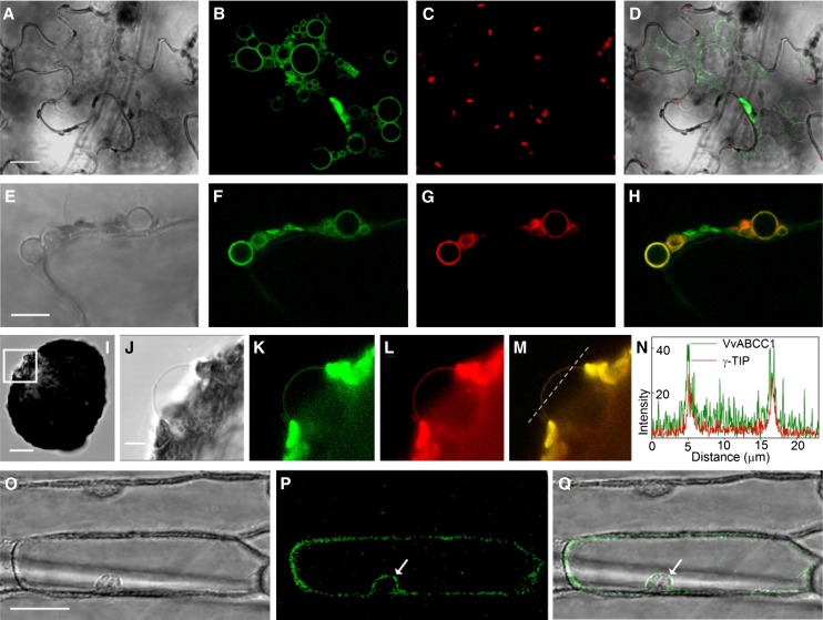
Figure 3.
ABCC1-GFP Localizes to the Tonoplast.
(A) Bright-field image of a tobacco epidermis cell transiently expressing ABCC1-GFP. Bar = 20 µm.
(B) GFP signal encircling small vacuoles, which only occur in tobacco epidermis cells overexpressing VvABCC1-GFP.
(C) Chlorophyll autofluorescence corresponding to the image in (E).
(D) Bright-field, chlorophyll, and GFP fluorescence overlay image.
(E) Bright-field image of the VvABCC1-GFP overexpression-induced small vacuoles in a tobacco epidermis cell. Bar = 10 µm.
(F) VvABCC1-GFP signal encircling small vacuoles.
(G) Signal of the tonoplast marker γ-TIP-RFP encircling small vacuoles.
(H) Overlay image of VvABCC1-GFP and γ-TIP-RFP fluorescence showing colocalization of both dyes in small vacuole membranes.
(I) Bright-field image of a transiently transformed tobacco protoplast releasing a small vacuole (box). Bar = 20 µm.
(J) Close-up bright-field image of the small vacuole in (I). Bar = 5 µm.
(K) VvABCC1-GFP signal at the vacuolar membrane.
(L) Tonoplast marker γ-TIP-RFP at the vacuolar membrane.
(M) Overlay image of VvABCC1-GFP and γ-TIP-RFP fluorescence at the membrane encircling the released small vacuole.
(N) Fluorescence intensity over distance plot of VvABCC1-GFP and γ-TIP-RFP fluorescence along the dotted line in M.
(O) Bright-field image of onion epidermis cells transiently expressing VvABCC1-GFP. Bar = 50 µm.
(P) VvABCC1-GFP signal.
(Q) Bright-field and GFP fluorescence overlay image showing a signal around the nucleus indicative of tonoplast localization (arrows in [P] and [Q]).

