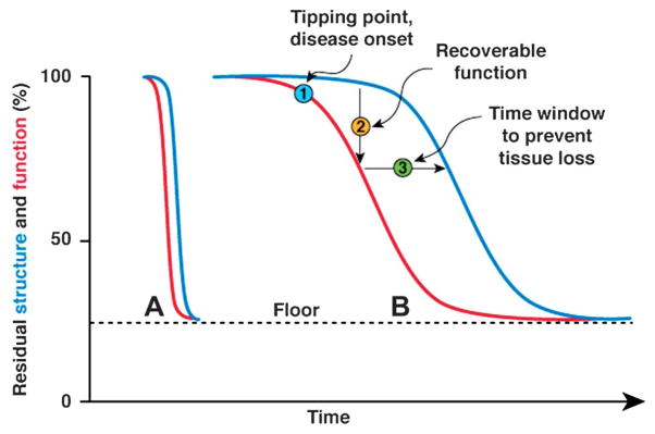
FIG. 2.

Conceptual model of structure–function relationship for a large population of retinal ganglion cells, in which different levels of cell dysfunction coexist. Condition A corresponds to a fast-progressing disease, such as Leber hereditary optic neuropathy. Condition B corresponds to a slow progressing disease such as glaucoma. The curves representing function and structure have identical form, although shifted by a time lag. Note that the residual function and structure measurable in advanced stages of the diseases with pattern electroretinogram or optical coherence tomography has a nonzero value (floor).
