
Figure 1.
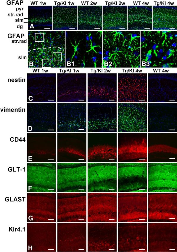
Progressive accumulation of astrocytic pathology in GFAPTg;Gfap+/R236H (Tg/KI) hippocampus at 1, 2, and 4 weeks of age compared with normal astrocyte development in wild-type (WT) mouse hippocampus. Immunostaining in A and B, GFAP; C, nestin; D, vimentin; E, CD44; F, GLT-1; G, GLAST; H, Kir4.1 immunostaining. A, Progressive increase of GFAP immunoreactivity, first in str.lac-mol (slm) and then in str. rad and str. mol is much more prominent in Tg/KI than in WT hippocampi. B, Layer-specific differences in astrocyte morphology in 2-week-old Tg/KI hippocampus. The border between slm and str.rad is dotted. Note that in str.rad (upper boxed area, enlarged in B1), astrocytes have only a slightly thickened appearance, whereas in the slm they have more complex profiles and many thin processes filled with GFAP (middle boxed area, enlarged in B2); in the outer part of the slm, many swollen astrocytes with thick proximal processes are present (lower boxed area, enlarged in B3). Confocal microscopy; pyr, pyramidal cell layer; str.rad, stratum radiatum; slm, stratum lacunosum-moleculare; dg, dentate gyrus. Scale bars: A, 130 μm; B, 70 μm; C–H, 110 μm.