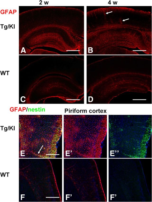
Figure 11.

Astrocyte changes are widespread in the neocortex of GFAPTg;Gfap+/R236H mice. Note the difference between levels of GFAP in neocortex in 2 and 4 week GFAPTg;Gfap+/R236H (A and B, respectively) mice versus age-matched WT mice (C and D, respectively). E, F, Reactive astrocytes populate the piriform cortex in GFAPTg;Gfap+/R236H mouse (E) and are absent from piriform cortex in WT mouse (F). Astrocytes around blood vessels (arrows, B, E) stained strongly for GFAP. Confocal microscopy; scale bars: A–D, 350 μm; E, F, 200 μm.
