
Figure 3.
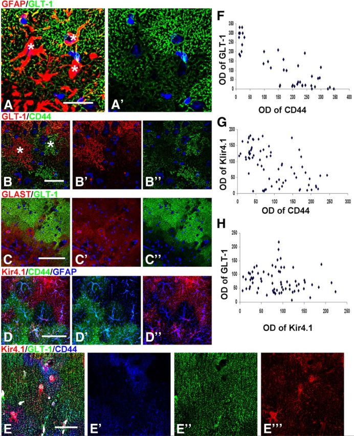
Microheterogeneity in immunohistochemical patterns of CD44, GLT-1, GLAST, and Kir4.1 in GFAPTg;Gfap+/R236H mouse hippocampus. A, Variability of GLT-1 immunostaining in adjacent astrocytes in the str. lac-mol. at 4 weeks; * denote locations of astrocyte nuclei (compare with Nissl); B, GLT-1 level is minimal in cells with high immunostaining for CD44; C, GLAST is present in some astrocytes without GLT-1 immunoreactivity. D, Focal diversity in the levels of CD44 and Kir4.1 immunoreactivity. Note that astrocytes with high CD44 immunoreactivity do not also have high Kir4.1 immunolabeling. E, Triple immunostaining for Kir4.1, GLT-1, and CD44 displays variable pattern of immunolabeling in neighboring astrocytes. Confocal microscopy; scale bars, 95 μm. F–H, Graphs representing optical density readings of CD44 versus GLT-1, CD44 versus Kir4.1, and Kir4.1 versus GLT-1 immunofluorescence, respectively. Note that inverse correlation is significant in F (strong: r = 0.854, p < 0.001) and G (weak: r = −0.573; p < 0.001); in H, there is no correlation between parameters (r = −0.156; p = 0.196).