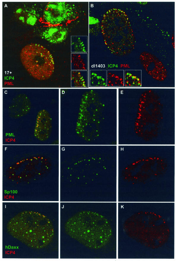
FIG.4.
Evidence for de novo formation of ND10-like structures in association with ICP4 complexes at the early stages of infection. (A) An HFFF-2 cell at the edge of a developing HSV-1 strain 17 plaque, showing ICP4 foci (green) commonly at the edge of the nucleus, some of which are in association with residual PML (red). The cells had been infected at an MOI of 0.001 and fixed and stained 24 h later, and then individual plaques were examined. (B) An HFFF-2 cell at the edge of a developing dl1403 plaque, showing extensive relocalization of PML into structures at the nuclear periphery. The cells had been infected at an MOI of 0.1 and fixed and stained 24 h later, and then individual plaques were examined. The lower cell is at an earlier stage of infection and shows a more normal distribution of PML. The insets in panels A and B show the separated and merged channels of a selected detailed area of each cell. (C to K) Evidence for de novo formation of ND10-like complexes containing PML, Sp100, and hDaxx in association with ICP4 complexes near the nuclear periphery at early times of high-multiplicity infections. HFFF-2 cells were infected with dl1403 at 10 PFU/cell and then stained for ICP4 (red; C to K) and PML (green; C to E), Sp100 (green; F to H), or hDaxx (green; I to K) 3 h after addition of the virus. The images show cells selected to illustrate the extensive recruitment of these three ND10 proteins into structures that are associated with ICP4 complexes near the nuclear periphery, in the absence of functional ICP0.

