
Figure 2.
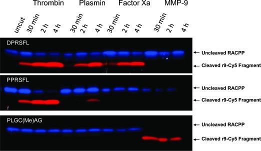
Peptide cleavage of RACPPs by purified thrombin, plasmin, factor Xa, and MMP-9. Three RACPPs (top: DPRSFL, middle: PPRSFL, and bottom: PLGC(Me)AG) were separately exposed to purified enzymes for the times indicated. Peptide cleavage products were separated by electrophoresis using tricine-SDS polyacrylamide gels and imaged using the Maestro with 620 nm excitation, and emission collected for Cy5 (660 to 720 nm) or Cy7 (760 to 830 nm). Ratiometric images were produced by dividing the Cy5 emission with Cy7 emission and pseudocolored from blue (ratio minimum) to red (ratio maximum).