FIGURE 4:
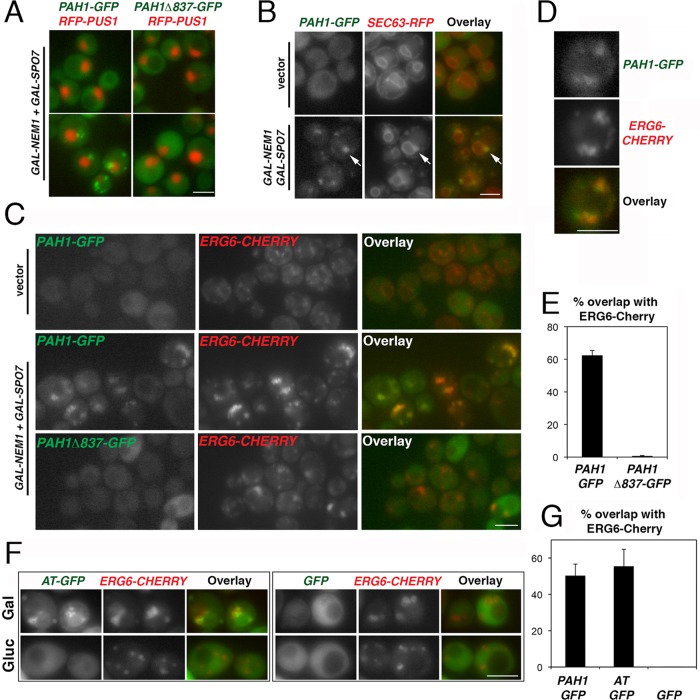
The acidic stretch is necessary and sufficient for the Nem1p-Spo7p–directed recruitment of Pah1p-GFP to lipid droplets. (A) pah1Δ cells expressing the intranuclear reporter RFP-PUS1 and either PAH1-GFP (left) or PAH1Δ837-GFP (right) were transformed with GAL-NEM1 and GAL-SPO7, transferred from raffinose- to galactose-containing medium, grown for 6 h, and imaged by epifluorescence microscopy. Bar, 5 μm. (B) pah1Δ cells expressing PAH1-GFP and SEC63-RFP were transformed with either empty vectors (top) or GAL-NEM1 and GAL-SPO7 (bottom). Cells were grown in galactose-containing medium for 6 h as in A. Arrows point to typical ER-associated Pah1p-GFP labeling. Bar, 5 μm. (C) pah1Δ cells with chromosomally integrated ERG6-Cherry expressing PAH1-GFP (top and middle) or PAH1Δ837-GFP (bottom) were transformed with either empty vectors (top) or GAL-NEM1 and GAL-SPO7 (middle and bottom). Cells were grown in galactose for 6 h as in A. (D) Representative PAH1-GFP cell overexpressing GAL-NEM1 and GAL-SPO7 from C. (E) The percentage of cells in which Pah1p-GFP or Pah1pΔ837-GFP overlaps with more than half of Erg6p-Cherry puncta per cell is given. Values represent the mean ± SD of three independent experiments. At least 300 cells per experiment and strain were scored. (F) Nem1p-Spo7p–dependent targeting of the Pah1p acidic stretch to lipid droplets. The acidic tail fusion to GFP (AT-GFP, left) or GFP alone (right) was expressed under the control of the NOP1 promoter in RS453 cells coexpressing chromosomally integrated ERG6-Cherry. Cells were grown in galactose as in A (Gal) or glucose (Gluc) and imaged by epifluorescence microscopy. Bar, 5 μm. (G) RS453 ERG6-Cherry cells expressing PAH1-GFP, AT-GFP, or GFP, all under the control of the NOP1 promoter, were grown in galactose as in F, and the percentage of overlap between the GFP fusions and Erg6p-Cherry was quantified as described in E.

