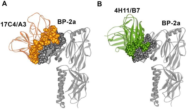
Figure 4. Molecular mAbs/BP-2a 515 variant docking reveals two different binding orientations.
(A) Ribbon representation of mAb 17C4/A3-BP-2a 515 variant complex. (B) Ribbon representation of mAb 4H11/B7-BP-2a 515 variant complex. In both complexes, the binding interface is CPK represented and colored according to the mAbs and antigen overall structure.

