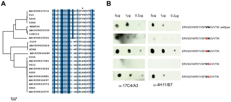
Figure 7. Single amino acid contribution to mAbs-antigen interaction.
(A) Sequence alignment of the identified epitope region in the 22 BP-2a allelic variants. The alignment is colored according to sequence identity from dark blue to white. The neighbor-joining phylogenetic tree has been constructed with the full 22 BP-2a protein sequences. The epitope alignment clearly illustrates a mosaic structure in the protein indicating the presence of recombination events. An asterisk indicates a position under positive selection. (B) Peptide dot blot immune assay of wild type and mutated peptide with 17C4/A3 and 4H11/B7 mAbs. The mutated residues within each synthetized peptide are highlighted in red.

