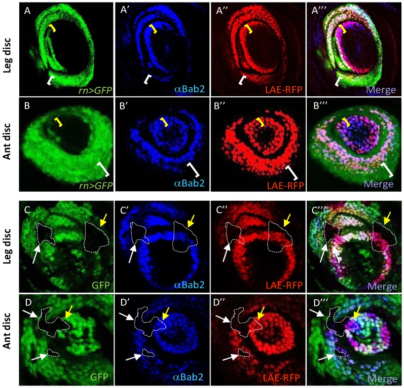
Figure 6. rotund is required for bab2 activation only in proximal-most rings, in both leg and antenna.
GFP expression (green), Bab2 immunostaining (blue) and LAE-RFP expression (red) alone, as well as the merged images of leg (A–A″′ and C–C″′) and antennal (ant) (B–B″′ and D–D″′) discs from late third-instar larvae, either wild-type (A–B) or carrying rn null mutant clones (C–D), are shown. rn expression was monitored by combining rn-Gal4 and UAS-GFP constructs (rn>GFP). LAE regulatory activity was monitored with a LAE-RFP construct (Materials and methods). rn-Gal4 reporter expression is not detected in the distal-most bab2-expressing cells (yellow brackets), and extends more proximally than bab2 expression (white brackets). In mosaic tissues, rn16 mutant clones (examples are circled by dashed lines) are detected by the absence of GFP (black areas). Note that only proximally-located mutant cells (white arrows), and not those located in the distal-most ring (yellow arrows), display strongly diminished bab2 and LAE-RFP expression, in either leg or antennal tissues.

