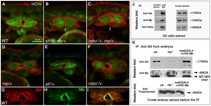
Figure 2. Genetic and physical association between Mp, Slit and Robo.
Cardiac cross sections of wild type (A), slit/+;mp/+ (B), robo/+;mp/+ (C) double heterozygous, and mp/+ (D), slit/+ (E), robo/+ (F) single heterozygous labeled with anti HOW (green), and anti Dg (red). The cardiac lumens (marked by arrowheads) of the double-heterozygous mutant are smaller relative to the control. G-I: cardiac cross sections of wild type embryos labeled with anti Slit (G,I red) and anti Mp (H,I green), indicating their co-localization along the lumen. J- immunoprecipitation with anti Slit antibodies (or with a control normal mouse serum) of an extract of S2 cells co-transfected with Slit, Robo, and Mp. The same blot was then reacted individually with anti- Slit, Mp, and Robo corresponding to the three upper lanes). The anti Mp antibody reacted with a single band of ∼120 kDa, corresponding to mp cDNA 3hnc1. This immunoprecipitation (IP) is representative of three independent IP experiments. The crude extracts contained comparable amounts of transfected proteins as indicated by the antibody reactivity with each of the transfected cDNA constructs presented in the right panel. K-Immunoprecipitation with anti Slit antibodies of comparable protein extracts taken from stage 16 control (yw), mp−/−, or embryos overexpressing Mp in heart and muscles (using mef2-GAL4 driver). Western blot with anti Slit of the IP material shows elevated levels of Slit in the Mp-overexpressing embryos and reduced Slit levels in mp mutants. Reaction of the same blot with anti Mp antibodies (lower panel) revealed a specific band of ∼39 kDa, corresponding to the Endostatin fragment. Western blot with anti Tropomyosin of the embryo protein extracts before taken to the IP with Slit, is shown in the lower panel, indicating comparable protein levels in each of the samples.

