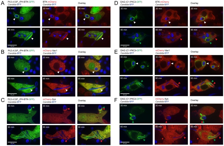
Figure 5. BTK-mCherry and mCherry-Vav1 colocalize with PI(3,4,5)P3 but not with DAG.
(A): Colocalization of BTK-mCherry and PH-BTK-GFP that binds to PI(3,4,5)P3 at 30 and 90 minutes of coincubation with Candida-BFP showing phagocytosis of yeast and hyphae, respectively. (B): Colocalization of mCherry-Vav1 and PH-BTK-GFP at 30 and 90 minutes of coincubation with Candida-BFP. (C): Localization of mCherry-Syk and PH-BTK-GFP at 30 and 90 minutes of coincubation with Candida-BFP. (D): Localization of BTK-mCherry and C1-PKCδ-GFP at 30 and 90 minutes of coincubation with Candida-BFP. (E): Localization of mCherry-Vav1 and C1-PKCδ-GFP at 30 and 90 minutes of coincubation with Candida-BFP. (F): Localization of mCherry-Syk and C1-PKCδ-GFP at 30 and 90 minutes of coincubation with Candida-BFP. White arrows indicate areas of co-localization, while red and green arrows indicate areas of speciation. Experiments were performed at least three times, representative micrographs are shown.

