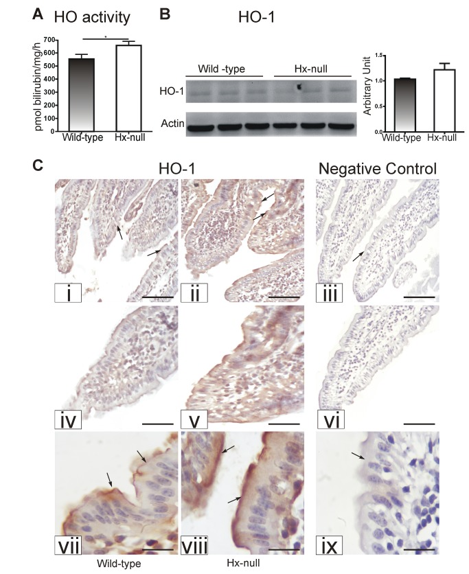
Figure 4. Hx deficiency results in enhanced heme catabolism in the duodenum.
(A) HO activity in the duodenum of wild-type and Hx-null mice. Data represent mean ± SEM; n= 8 for each genotype. * = P<0.05. (B) Representative Western blot of HO-1 expression in the duodenum of wild-type and Hx-null mice. Band intensities were measured by densitometry and normalized to actin expression. Densitometry data represent mean ± SEM; n=3 for each genotype. (C) Sections of the duodenum of a wild-type mouse (i, iv, vii) and an Hx-null mouse (ii, v, viii) stained with an antibody to HO-1. Enlarged details of sections i, ii, iii are shown in iv, v, vi respectively The HO-1-positive signal was more intense in the Hx-null mouse than in the wild-type control (arrows) Sections on the right (iii, vi, ix) represent negative controls in which the primary antibody was omitted. Bar i, ii, iii = 100µm; bar iv, v, vi = 57 µm; bar vii, viii, ix = 20 µm.

