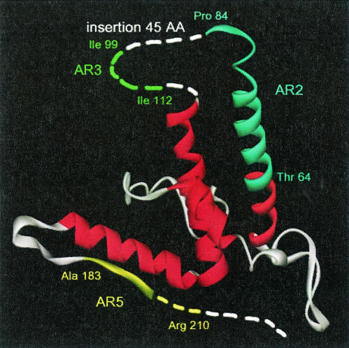
FIG. 6.
3-D model of DHBV core protein. The model was built as described in Materials and Methods with 1GQT as the molecular template (33). All helices have been stained in red. Three identified ARs have been positioned onto the 3-D model (AR2 from amino acids 64T to 84P is indicated in blue; AR3 from aa 99I to 112I is indicated in green in the region with unavailable atomic coordinates, corresponding to the 45-aa insertion; and AR5 from aa 183A to 210R is indicated in yellow). The dotted lines indicate regions in DHBc for which no atomic coordinates are available.

