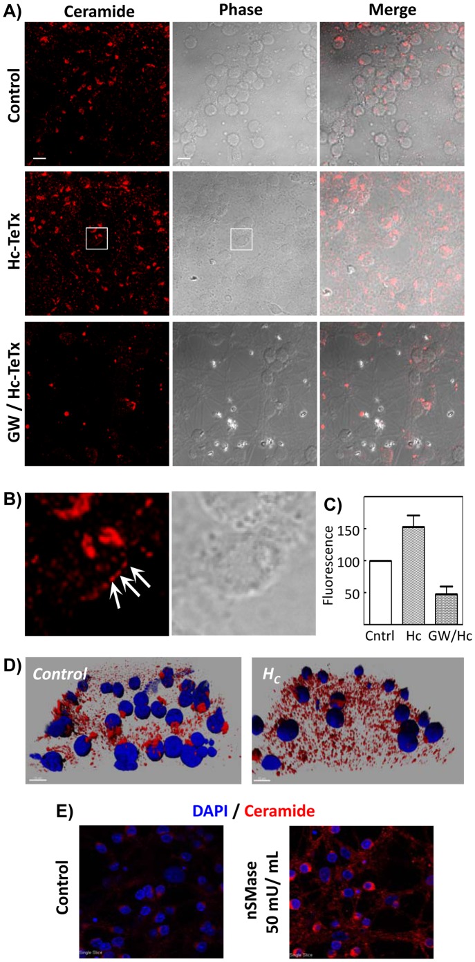
Figure 3. Hc-TeTx induces the increase of ceramide-platform content in CGN, due to nSMase action.
A) Cells were treated with Hc-TeTx 10 nM for 30 min, with or without pretreatment with 20 µM GW4869 for 1 h. Control cells were left totally untreated. After treatment cells were fixed, stained with anti-ceramide-antibody (clone 15B4) and analyzed by confocal microscopy. As can be seen in A, the results indicate the formation of ceramide-enriched membrane platforms upon Hc treatment, which is prevented by nSMase inhibition with GW4869. Bright field images and merges are also shown. The images are representative for each three independent experiments. Bars in the control images represent 100 µm. B) Magnifications of the areas indicated in the Hc-treated cultures shown in section A. Ceramide clusters in the plasma membrane are indicated by arrows. C) Quantitative analysis of the formation of ceramide-enriched membrane platforms upon Hc treatment, which indicates nSMase-dependent increase in ceramide-platforms. The assessment of the fluorescence was performed using the Advanced Fluorescence Lite software from Leica. The fluorescence from 18 z planes was measured to obtain every determination. For every condition, 10 fields were measured with a mean of 25 cells per field. The results represent the mean ± SD of three independent studies. D) Tridimensional composition of cells without or with treatment with Hc-TeTx 10 nM for 30 min. The nuclei were stained with DAPI (blue) and ceramide was detected with the 15B4 antibody and a secondary antibody coupled to Alexa 546 (red). Fluorescence from 18 z planes was captured by confocal microscopy and tridimensional images were created with Imaris 6.3.1 software. E) CGN were treated or not with 50 mU/mL of nSMase from Bacillus Cereus for 30 min and subsequently labeled with the anti-ceramide antibody (red) and with DAPI (nuclei).

