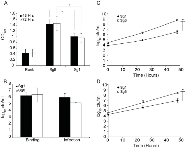
Figure 3. Intracellular growth of L. pneumophila.
(A) Biofilm production by Sg1 and Sg6 (crystal violet staining, OD600 nm). (B) Binding and invasion of Sg6 str. Thunder Bay compared to Sg1 str. Philadelphia to/within human NCI-H292 lung epithelial cells. (C) Intracellular replication of L. pneumophilla in A. castellani. The magnitude of replication is reported in log10 CFU/ml. (D) Intracellular growth of Sg6 str. Thunder Bay (square) compared to Sg1 str. Philadelphia (diamond) within U937 derived Human Macrophage cells. Each data point is an average of three independent experiments. For each experiment data was collected from 3 wells and a mean value was determined. *denotes statistical significance as determined by a two-tailed student’s t-test with a P-value <0.05.

