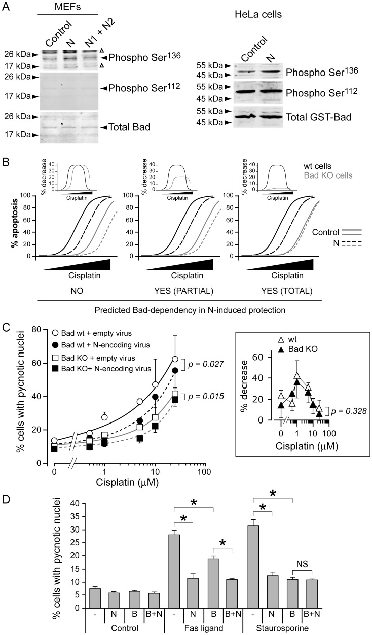
Figure 2. Bad modulation plays little role in fragment N-induced cell survival.
A. MEFs were infected with an empty lentivirus or with lentiviruses encoding fragments N, N1, and N2, as indicated. Alternatively, HeLa cells were transfected with a plasmid encoding a GST-Bad fusion protein with either empty pcDNA3 or with pcDNA3 encoding fragment N. The cells were lysed 24 hours later. The extent of Bad phosphorylation was assessed by Western blot using antibodies specific for Bad phosphorylated on serine 112 or serine 136. B. Predicted apoptotic response in cells lacking or not Bad in the presence or in the absence of fragment N. See text for details. C. MEFs expressing or not Bad were infected with an empty lentivirus or with a fragment N-encoding lentivirus. Forty-eight hours later, the cells were incubated with the indicated concentrations of cisplatin. Apoptosis was assessed after an additional 24 hour-period. The inset depicts the reduction in the percentage of apoptosis induced by the expression of fragment N in wild-type or Bad KO cells at the indicated doses (using the values of the main figure). The results correspond to the mean ±95% CI of sextuplets derived from 4 independent experiments. Statistics were done by repeated measures ANOVA tests. D. MEFs were infected with an empty lentivirus (-), with a Bcl-2 (B)-encoding lentivirus, and with a fragment N (N)-encoding lentivirus, in the indicated combinations. Fourty-eight hours later, the cells were incubated with 30 ng/ml Fas ligand or 10 nM staurosporine. Apoptosis was assessed after an additional 24 hour-period. The results correspond to the mean ±95% CI of 4–8 independent experiments. Asterisks denote statistically significant differences [one-way ANOVAs followed by pair-wise Dunn (Bonferroni) post hoc t tests]; NS, not significant.

