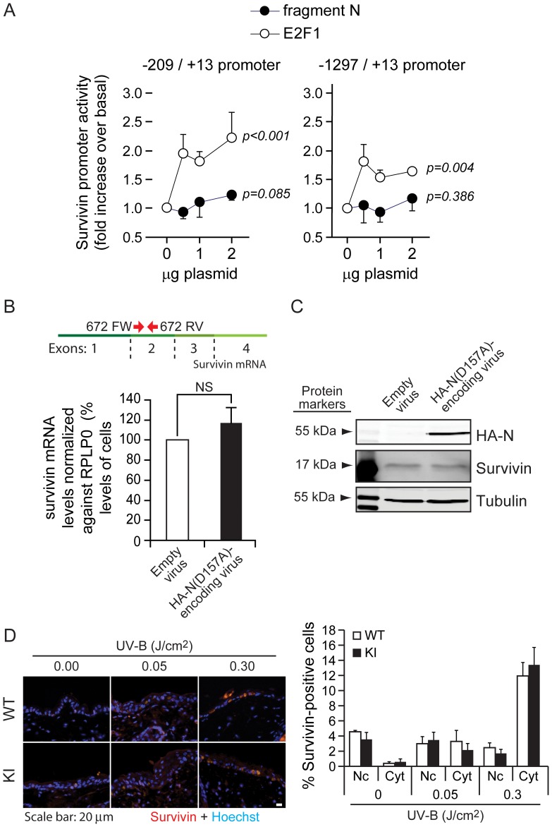
Figure 3. Fragment N does not regulate survivin expression.
A. MIN6 cells were co-transfected with a luciferase expressing vector under the control of either a minimal promoter sequence allowing the transcription of survivin (left graph) or the entire survivin promoter sequence (right graph) with increasing amounts of fragment N- (closed circles) or E2F1- (open circles) encoding plasmids. The results correspond to the mean ±95% CI of 6 (left panel) and 3 (right panel) independent experiments performed in triplicate. Repeated measures ANOVA tests were performed to determine if there was a significant increase in luciferase activity induced by the E2F1- or fragment N-encoding plasmids (normality was verified with the Shapiro-Wilk test). B–C. MEFs were infected with an empty virus or with a lentivirus encoding the HA-tagged version of the D157A fragment N mutant. Survivin mRNA levels were analyzed 24 hours later by quantitative RT-PCR (panel B). The location of the 672FW and 672RV primers (red arrows), used for the amplification of the survivin mRNA, is depicted on top of the graph. Alternatively, cells were lysed and the protein expression of HA-fragment N and survivin was assessed by Western blotting (panel C). The results correspond to the mean ±95% CI of 3 independent experiments. D. Skins of mice were irradiated with low (0.05 J/cm2) and high (0.3 J/cm2) doses of UV-B light. Expression of nuclear and cytoplasmic survivin was assessed by immunofluorescence in situ (left panel). The quantitation shown on the right-hand side corresponds to percentages of keratinocytes displaying nuclear or cytoplasmic survivin (mean ±95% CI of 6 and 10 mice for the low and high UV-B dose exposure, respectively). No cells were found to display both cytoplasmic and nuclear survivin expression.

