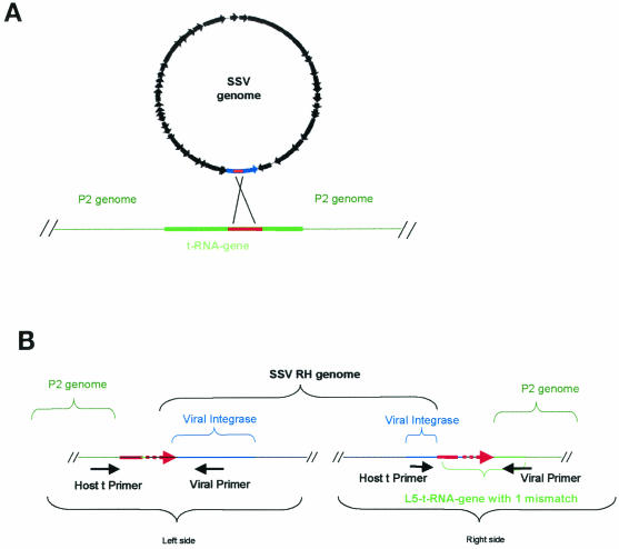
FIG. 3.
Integration of SSV RH and SSV K1 into the S. solfataricus P2 genome. (A) General overview of SSV integration into a tRNA gene of S. solfataricus P2. The circular SSV genome is in black, the blue region represents the virus-encoded integrase, the green lines indicate host chromosome sequences, and the solid red lines indicate putative attP and attA sites in the virus and the host. (B) Schematic of SSV RH integrated into the host chromosome at the L5 tRNA gene. The general locations of viral integration and host primers that were used in the PCR-based integration assay are shown directly below the genomes. The dashed red lines indicate the region within which the recombination event occurs.

