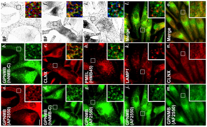
Figure 1. Localization of GPNMB within melanocytic MNT-1 cells by IFM.
MNT-1 cells were fixed with methanol, labeled with the indicated primary antibodies and fluorochrome-conjugated species-specific secondary antibodies, and analyzed by IFM-D (a, b, d, e, g, h, j–o) and by bright field microscopy (c, f, i). a–c, anti-GPNMB antibodies AF2550 and hNMB-C; d–f, hNMB-C and anti-calnexin (CLNX); g–i, AF2550 and anti-PMEL antibody HMB45; j–l, AF2550 and anti-LAMP1 antibody (H4A3); m–o AF2550 and CLNX. Boxed regions are magnified 4X in the insets at bottom right of each panel; merged images are shown in the insets of panels c, f, i, l and o, and in a–f, melanosomes (from bright field images) are pseudocolored blue. Bar, 10 μm.

