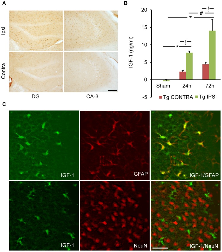
Figure 1. Trauma-induced astrocyte-specific IGF-1 overexpression.
A) Coronal sections from IGF-1Tg mice show a marked increase in IGF-1 immunolabeling in the hippocampal dentate gyrus (DG) and CA-3 regions in the ipsilateral (ipsi) hemisphere compared to the contralateral (contra) after lateral controlled cortical impact (CCI) brain injury. Immunohistochemical staining was performed using an antibody that detects both human and mouse IGF-1. Representative picture is taken from an IGF-1Tg mouse 72 h after CCI. Scale bar = 100 µm. B) Quantification of IGF-1 expression using a human-specific IGF-1 ELISA. IGF-1 concentrations increased progressively in the ipsilateral hippocampus of IGF-1Tg mice at 24 h and 72 h after severe CCI brain injury. Data plotted as mean+SEM. * p<0.05 comparing injured with sham, ! p<0.05 comparing ipsilateral to contralateral, and # p<0.05 comparing 72 h to 24 h. C) Confocal images taken from ipsilateral hemisphere bordering the contused cortex demonstrate widespread colocalization of IGF-1 (green) with the astrocyte-specific marker GFAP (red) in brain-injured IGF-1Tg mice. No co-localization of IGF-1 (green) and neuron-specific NeuN (red) was observed. Scale bar = 50 µm for C.

