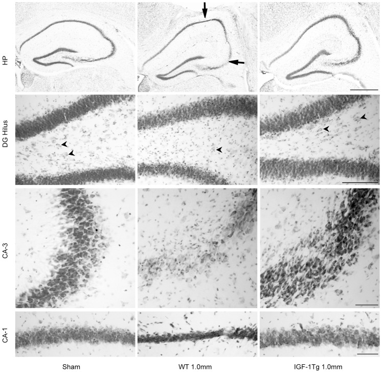
Figure 6. IGF-1 overexpression promoted hippocampal neuronal survival at 72 h after severe brain injury.
Representative images of the ipsilateral hippocampus (HP) from Nissl-stained brain sections illustrate pallor or thinning of CA-3 and CA-1 areas of pyramidal layer (arrows) in wildtype (WT) mice. Higher magnification images from DG, CA-3 and CA-1 areas demonstrate hilar and CA-3 neuronal loss and thinning of the CA-1 pyramidal layer. IGF-1 overexpressing (IGF-1Tg) mice showed marked neuroprotection in each hippocampal subregion. Arrowheads point to hilar neurons. Scale bars = 500 µm (top HP panel), 100 µm for DG panel, 50 µm for CA-3 and CA-1 panels.

