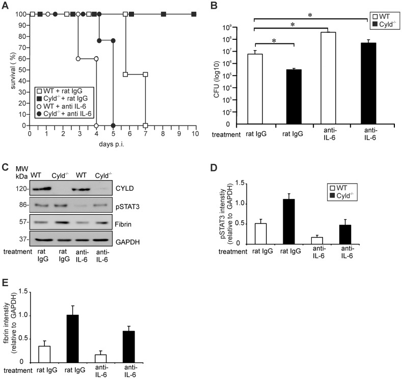
Figure 6. IL-6 neutralization abolishes increased STAT3 activation, fibrin production, survival and pathogen control in Lm-infected Cyld−/− mice.
(A) The survival rates of rat IgG and α-IL-6-treated Lm-infected WT and Cyld−/− mice (n = 7 per experimental group) are shown. The survival rate of IgG-treated Cyld−/− mice (p<0.05) but not of the other groups was significantly increased as compared to rat IgG-treated WT mice. (B) CFUs were determined in the liver of Lm-infected rat IgG and IL-6-treated WT and Cyld−/− mice at day 5 p.i. (n = 5 per experimental group; * p<0.05). Data show the mean ± SD from one of two representative experiments. (C) Proteins were isolated from livers of infected rat IgG and IL-6-treated WT and Cyld−/− mice (n = 3 per experimental group) at day 5 p.i. WB analysis for CYLD, pSTAT3, fibrin and GAPDH was performed and representative data are shown. (D, E). Quantification of hepatic pSTAT3 (D) and fibrin (E) (± SD) was performed from WB data of rat IgG and α-IL-6-treated WT and Cyld−/− mice, respectively. The results present 3 mice of each experimental group.

