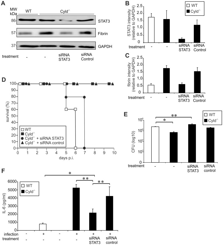
Figure 7. Inhibition of STAT3 reduces fibrin production, survival and pathogen control in Lm-infected Cyld−/− mice.
(A) Proteins were isolated from infected livers of untreated WT and Cyld−/− mice as well as STAT3 siRNA and control siRNA-treated Cyld−/− mice at day 5 p.i. (n = 3 experimental group). WB analysis for CYLD, pSTAT3, fibrin and GAPDH was performed and representative data are shown. (B, C) Quantification of total STAT3 (B) and fibrin (C) (± SD) was performed from WB data of livers from Lm-infected WT and Cyld−/− mice, which were treated as indicated. The results present n = 3 mice per experimental group. (D) The survival rates of infected WT and Cyld−/− as well as STAT3 siRNA and control siRNA-treated Cyld−/− mice are shown. The survival of Cyld−/− and control siRNA-treated Cyld−/− mice but not of STAT3 siRNA-treated Cyld−/− mice was significantly increased as compared to WT animals (p<0.05 for both groups, n = 5 per experimental group). Survival was monitored until day 10 p.i. One of two representative experiments is shown. (E) CFUs were determined in the liver of Lm-infected untreated WT and Cyld−/− mice as well as STAT3 siRNA and control siRNA-treated Cyld−/− mice at day 5 p.i. (* p<0.05, ** p<0.01; n = 5 per experimental group). Data show the mean ± SD and one of two representative experiments. (F) The serum concentration of IL-6 was determined by a cytometric bead assay at day 5 p.i. Data show the mean + SD of 5 mice per experimental group and from one of two representative experiments (* p<0.05, ** p<0.01).

